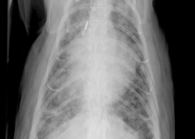
Brutus is a 13 year old male neutered Domestic Short Haired cat.
Brutus has a history of coughing for 3-4 weeks. He has lost some weight and stopped eating ~3 days ago. On physical examination he is thin, with wheezes and crackles auscultated over all lung fields.
Thoracic radiographs were made:
- What type of lung pattern does Brutus have?
- What are your differential diagnoses?
Answers
There is a severe increase in bronchial markings (ie increased “dough-nuts”) throughout all lung lobes. The lungs have an overall increase in soft tissue opacity (they are too white) and the vessels are hazy and hard to define as a result. In places, it appears that bronchial markings are bunched together as nodules, particularly in the left caudodorsal lung on the VD projection. The diaphragm on the left lateral projection is fairly straight. On the ventrodorsal radiograph, the left and right thoracic walls are convex relative to the heart, making the thorax appear widened. These changes are consistent with moderately hyperinflated lungs. During the examination, Brutus was tachypnoeic.
Radiographic conclusion: diffuse, severe bronchointerstitial lung pattern which is coalescing to a nodular lung pattern in places.
The most likely differential for these severe changes is diffuse pulmonary neoplasia (bronchial carcinoma, possibly metastatic carcinoma). Abdominal ultrasound examination was unremarkable. Fine needle aspirates of the lungs were made with ultrasound guidance, and were not diagnostic. Bronchial carcinoma was diagnosed after post-mortem histopathological examination.


